
The Dixon fat-suppression technique, first proposed by Dixon, is based on the chemical shift difference between water and fat. By using different echo times, it acquires signals from water and fat protons in both in-phase and opposed-phase conditions.
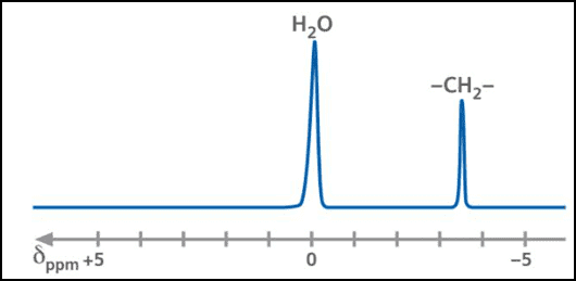
When water and fat signals are in phase, the collected signal is:
S1 = W + F
When water and fat signals are opposed in phase, the collected signal is:
S2 = W – F
By adding the two signals:
S1 + S2 = 2W
This eliminates the fat component, producing a pure water-proton image—achieving fat suppression.
By subtracting the signals:
S1 – S2 = 2F
This yields a pure fat signal, generating a fat image. This is the original two-point Dixon method, which has since been extended into multiple variations, including the three-point Dixon technique.
Applications of the Dixon Technique
The Dixon method is widely applied in studies of liver fat, obesity and metabolic disorders, fatty tumours, renal imaging, and focal hepatic lesions, among others.
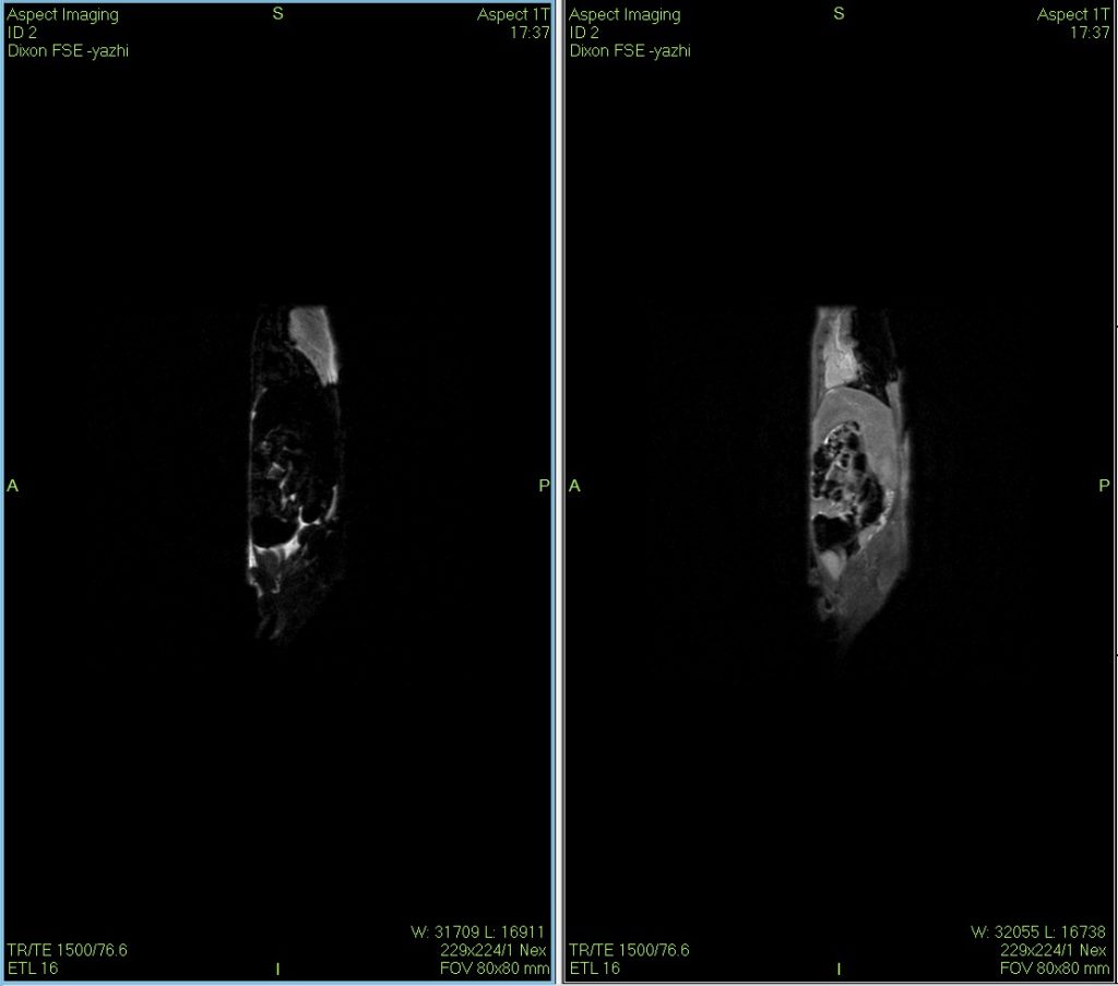
Dixon water–fat imaging of a mouse using MRI
The Dixon water–fat separation technique is a versatile MRI method with unique value in diagnosing, differentiating, and evaluating therapeutic responses in certain animal disease models.
 Scan QR Code
 Scan QR Code
Phone: 400-060-3233
After-sales: 400-060-3233


Back to Top