mohoso
Imágenes de animales pequeños Métodos
Métodos de obtención de imágenes de animales pequeños
Imágenes de animales pequeños Se refiere al uso de diversas técnicas de imagen para estudiar y visualizar la anatomía., fisiología, y patología de pequeños animales de laboratorio., típicamente roedores como ratones y ratas. Estas técnicas de imagen permiten a los investigadores examinar y controlar de forma no invasiva la progresión de la enfermedad., evaluar la eficacia del tratamiento, y estudiar procesos biológicos en animales vivos..
Comúnmente se emplean varias modalidades de imágenes en imágenes de animales pequeños:
- Imagen de resonancia magnética (resonancia magnética): La resonancia magnética utiliza fuertes campos magnéticos y ondas de radio para generar imágenes detalladas de las estructuras internas de los animales.. Proporciona un excelente contraste de tejidos blandos y permite a los investigadores visualizar características anatómicas., detectar tumores, y estudiar la función de los órganos.
- Tomografía computarizada (Connecticut): La tomografía computarizada implica tomar múltiples imágenes de rayos X desde diferentes ángulos y combinarlas para crear imágenes transversales detalladas del cuerpo del animal.. La TC proporciona imágenes de huesos de alta resolución, vasos sanguineos, y organos, y es particularmente útil para estudiar anomalías esqueléticas y enfermedades vasculares..
- Tomografía de emisión de positrones (MASCOTA): Las imágenes PET utilizan la inyección de un trazador radiactivo que emite positrones.. Los positrones emitidos chocan con los electrones., produciendo rayos gamma que son detectados por el escáner PET. Esta técnica permite a los investigadores rastrear y cuantificar los procesos metabólicos., estudiar objetivos moleculares específicos, y evaluar la distribución de medicamentos radiomarcados en el cuerpo del animal.
- Tomografía computarizada por emisión de fotón único (ESPECTACULAR): Las imágenes SPECT son similares a las PET, pero utiliza diferentes trazadores radiactivos que emiten fotones individuales. SPECT proporciona imágenes en 3D de la distribución del radiotrazador en el cuerpo, permitiendo la evaluación de procesos fisiológicos y la detección de objetivos moleculares específicos.
- Imagen óptica: Técnicas de imagen óptica., tales como imágenes de bioluminiscencia e imágenes de fluorescencia, utilizar la luz para visualizar moléculas o células específicas dentro del animal. Las imágenes de bioluminiscencia implican la detección de luz emitida por reporteros bioluminiscentes., mientras que las imágenes de fluorescencia utilizan sondas fluorescentes que emiten luz cuando se excitan con una longitud de onda específica. Estas técnicas se utilizan ampliamente para rastrear la expresión genética., estudiando procesos celulares, y seguimiento del crecimiento tumoral en animales pequeños.
- Imágenes por ultrasonido: El ultrasonido utiliza ondas sonoras de alta frecuencia para crear imágenes de estructuras internas en tiempo real.. Es particularmente útil para obtener imágenes del sistema cardiovascular., visualizando tumores, y guiar las intervenciones en pequeños animales.
Imágenes de animales pequeños juega un papel crucial en la investigación preclínica, Permitir a los investigadores obtener información sobre los mecanismos de la enfermedad., evaluar nuevas terapias, y desarrollar nuevas herramientas de diagnóstico. Permite estudiar la progresión de la enfermedad en tiempo real., Reduce el número de animales necesarios para los experimentos., y proporciona una alternativa no invasiva a los métodos invasivos tradicionales.
Imágenes de animales pequeños por RMN de campo bajo
RMN de campo bajo (Resonancia magnética nuclear) se refiere al uso de tecnología de RMN con intensidades de campo magnético más bajas en comparación con los sistemas de resonancia magnética clínica convencionales.. Mientras que los sistemas de resonancia magnética de alto campo, normalmente operando en 1.5 tesla (t) o superior, Se utilizan comúnmente para imágenes humanas., Los sistemas de RMN de campo bajo funcionan con intensidades de campo que van desde unos pocos miliTesla (monte) a varias decenas de miliTesla (monte).
En el contexto de imágenes de animales pequeños, La RMN de campo bajo se puede utilizar para obtener información anatómica y funcional de pequeños animales de laboratorio.. Aquí hay algunos puntos a considerar con respecto a la aplicación de RMN de campo bajo en imágenes de animales pequeños:
Resolución y sensibilidad: Los sistemas de RMN de campo bajo generalmente proporcionan una resolución más baja en comparación con la resonancia magnética de campo alto. La resolución espacial de las imágenes puede verse limitada debido a la menor intensidad del campo magnético.. Sin embargo, para determinadas aplicaciones en la investigación con animales pequeños, como monitorear la progresión de la enfermedad o evaluar la respuesta al tratamiento, la resolución alcanzable aún puede ser suficiente.
Costo y accesibilidad: Los sistemas de RMN de campo bajo suelen ser menos costosos y más fáciles de mantener en comparación con los sistemas de RMN de campo alto.. Esto puede hacerlos más accesibles para investigadores con recursos limitados o aquellos que trabajan en laboratorios pequeños..
Consideraciones de seguridad: Los sistemas de RMN de campo bajo generalmente tienen intensidades de campo magnético más bajas., lo que puede dar lugar a menores problemas de seguridad en comparación con los sistemas de resonancia magnética de alto campo. Sin embargo, Sigue siendo importante seguir las pautas de seguridad y garantizar el manejo adecuado de los animales durante la toma de imágenes..
Aplicaciones: La RMN de campo bajo se puede emplear en varios imágenes de animales pequeños aplicaciones. Puede proporcionar información anatómica., como la morfología de los órganos, y puede usarse para monitorear la progresión de la enfermedad en estudios longitudinales. Además, La RMN de campo bajo se puede combinar con agentes de contraste o secuencias de pulsos específicas para extraer información funcional., como el flujo sanguíneo o la perfusión tisular, en animales pequeños.
Ventajas y limitaciones: El uso de RMN de campo bajo en imágenes de animales pequeños ofrece algunas ventajas, como la rentabilidad, facilidad de uso, y menores preocupaciones de seguridad. Sin embargo, la relación señal-ruido más baja (SNR) En comparación con la resonancia magnética de campo alto, puede limitar la sensibilidad de detección y la calidad general de la imagen.. Además, la disponibilidad de secuencias de imágenes especializadas y configuraciones de hardware puede ser más limitada en intensidades de campo magnético más bajas.
En resumen, La RMN de campo bajo puede ser una herramienta valiosa para imágenes de animales pequeños, ofreciendo capacidades de obtención de imágenes accesibles y rentables. Si bien puede tener limitaciones en términos de resolución y sensibilidad en comparación con la resonancia magnética de alto campo, aún puede proporcionar información anatómica y funcional valiosa para aplicaciones de investigación preclínica.
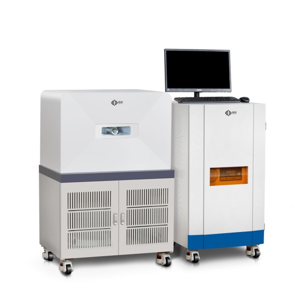
NIUMAG NM21-060H-I está diseñado para la observación de animales por resonancia magnética in vivo. Es un sistema de imanes permanentes que proporciona imágenes de alto contraste y tiene una interfaz de usuario intuitiva.. Como instrumento de resonancia magnética potente y no destructivo, Este sistema se utiliza ampliamente en ciencias biológicas para el estudio in vivo de estructuras tisulares y dispersión de agentes de contraste.. El sistema de resonancia magnética para animales pequeños de Niumag es robusto y tiene bajos costos de operación y mantenimiento..