NIUMAG
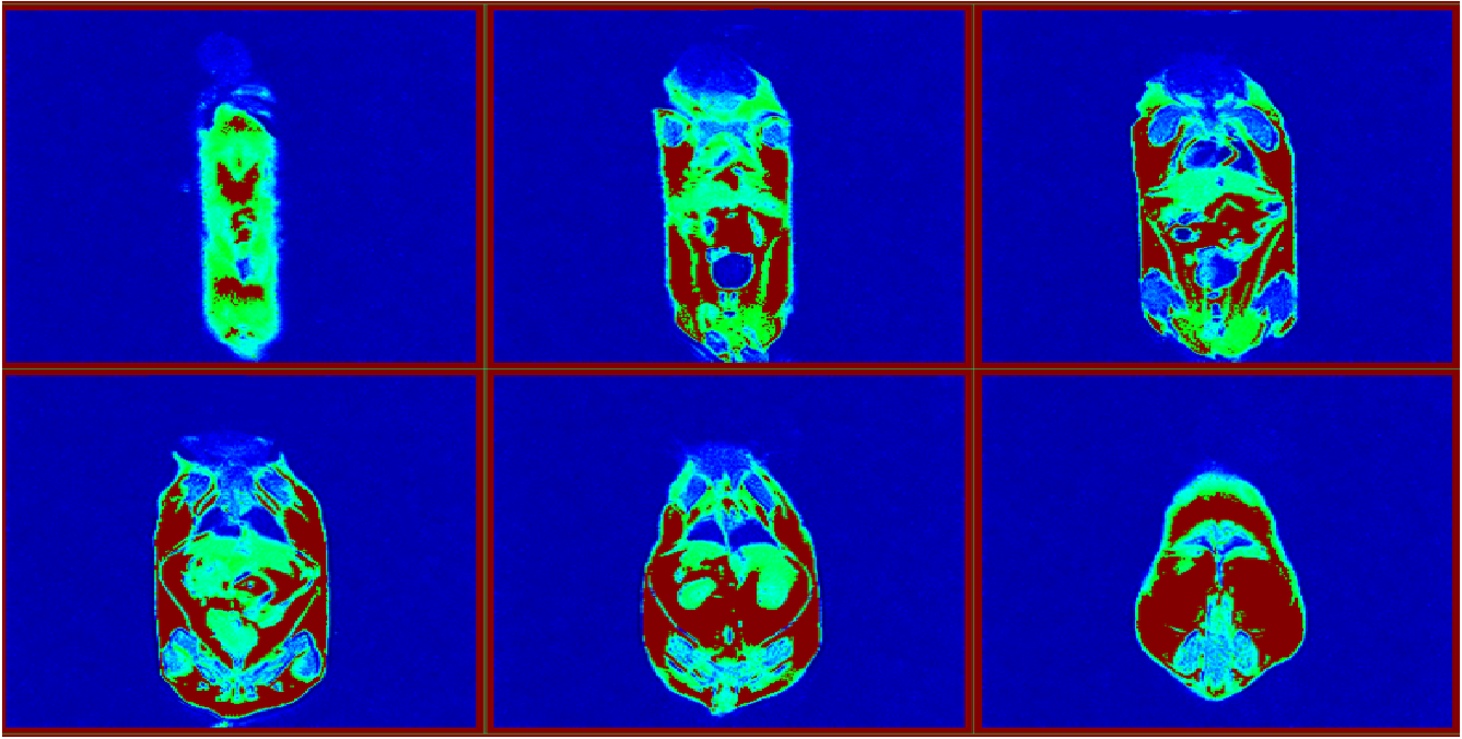
Mouse MRI by LF NMR
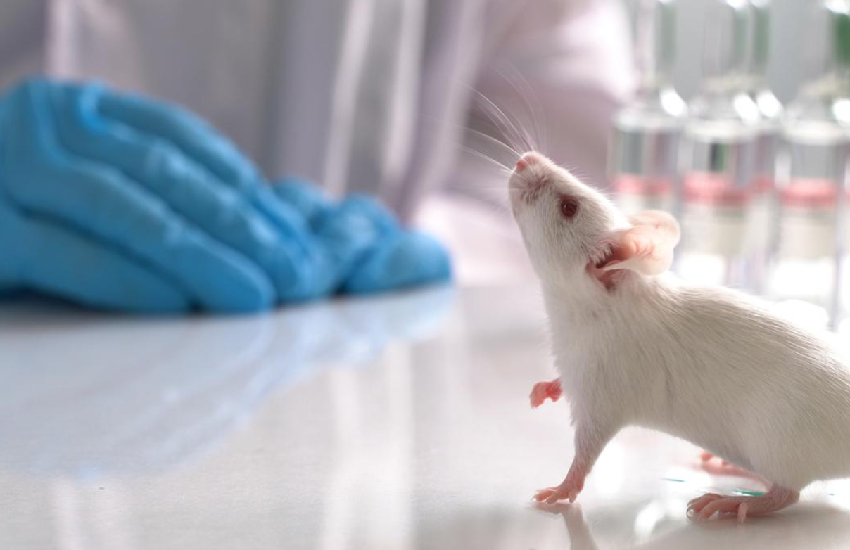
Mouse MRI (Magnetic Resonance Imaging) is a groundbreaking medical imaging technique that plays a pivotal role in research involving mice. By utilizing a powerful magnetic field, radio waves, and advanced computing, Mouse MRI produces intricate and comprehensive images of a mouse’s internal structures. This non-invasive method allows researchers to delve into the intricacies of organs, tissues, and other vital components within a living mouse.
Mouse MRI Appllication
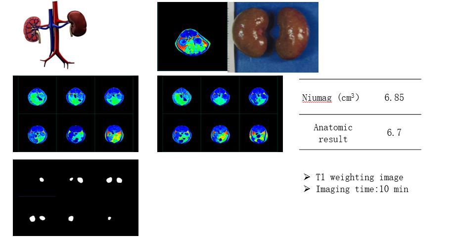
The applications of Mouse MRI are wide-ranging and immensely valuable. It enables the imaging of various structures, such as the brain, heart, lungs, liver, kidneys, and numerous other organs. Additionally, Mouse MRI proves indispensable in investigating disease processes, monitoring the effects of treatments, and evaluating drug efficacy.
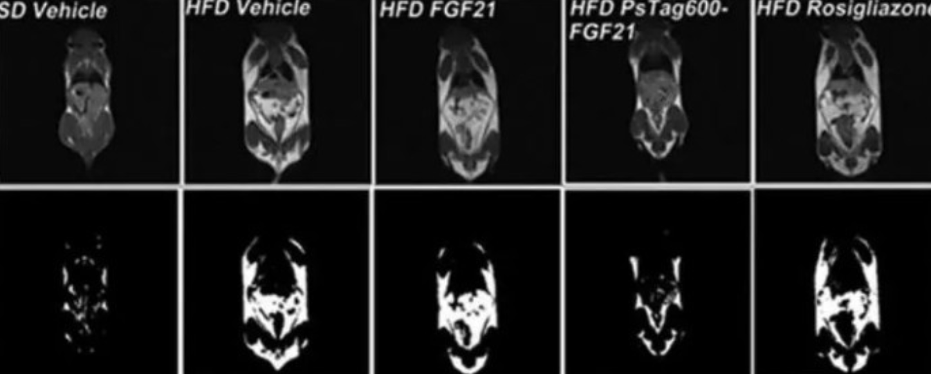
Different imaging techniques can be employed for Mouse MRI, each serving specific research purposes. T1-weighted imaging, T2-weighted imaging, diffusion-weighted imaging, and functional MRI are among the techniques that researchers may utilize based on the nature of their study.
Mouse MRI is especially significant for researchers investigating diseases and conditions that impact mice, such as cancer, Alzheimer’s disease, Parkinson’s disease, and cardiovascular ailments. Furthermore, it provides an avenue to study the effects of environmental toxins, drugs, and other treatments on the mouse body.
Mouse MRI and other Imaging Methods
There are several methods available to obtain images of mice for research purposes. Micro-Computed Tomography (micro-CT) utilizes X-rays to create high-resolution 3D images, primarily beneficial for studying bone structure, but capable of visualizing other tissues and organs as well. Optical imaging, utilizing light, allows researchers to study various biological processes, including gene expression and blood flow, through methods like bioluminescence imaging (BLI) and fluorescence imaging (FLI). Ultrasound, employing sound waves, is particularly useful for cardiovascular studies and examining blood flow. Positron Emission Tomography (PET) involves injecting a radioactive tracer into the mouse body and then detecting positrons emitted by the tracer using a PET scanner, which helps study metabolism and receptor binding. Similarly, Single Photon Emission Computed Tomography (SPECT) also employs radioactive tracers but uses a different type of tracer for studying a range of biological processes.
These methods, whether used individually or in combination, offer a comprehensive understanding of mouse anatomy, biology, and disease processes. They serve as invaluable tools for enhancing our knowledge of medical research and advancing treatments.
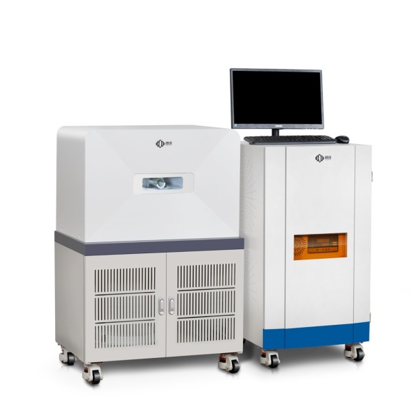
Mouse MRI by Low Field NMR
One promising technology for Mouse MRI is Low Field NMR (Nuclear Magnetic Resonance). Operating at a lower magnetic field strength than traditional clinical MRI machines, Low Field NMR holds significant potential in preclinical research involving mice.
The appeal of Low Field NMR lies in its affordability and accessibility, as it does not necessitate a specialized facility for operation. Moreover, it allows researchers to study a diverse array of mouse models and tissues, including the brain, liver, kidneys, and other vital organs.
The key advantage of Low Field NMR is its ability to study samples in their natural state, eliminating the need for invasive procedures. This is crucial for comprehending the natural structure and function of mouse tissues and organs, advancing our understanding of disease processes, and fostering the development of novel treatments.
Low Field NMR brings several advantages over traditional high field MRI machines. Its lower cost and reduced sensitivity to artifacts caused by movement or metal implants enhance image quality and make it an attractive option for researchers with budget constraints. Moreover, its decreased occurrence of motion artifacts is especially beneficial for studying organs like the mouse heart, which are prone to movement during imaging.
In conclusion, Mouse MRI, with its revolutionary techniques and evolving technologies like Low Field NMR, continues to shape the landscape of preclinical research and medical advancements.