NIUMAG
When performing a medical examination in a hospital, doctors often recommend that you do some necessary examination items, such as x-ray, CT, B-ultrasound, and nuclear magnetic resonance. So, what is the difference between x-ray, CT, B-ultrasound and nuclear magnetic resonance, and when is it appropriate to do it? This article tells you how X-ray, CT, B-ultrasound and MRI can play different roles.
X-ray: like squeezing bread
X-rays will pass through the human body and when they encounter the blocked part, they will not be exposed on the film. After the film is rinsed, this part will be white.
Just like a piece of bread or a piece of cotton, you cannot see the fiber structure inside, but if you press it with your hand, it will be clearer. The biggest disadvantage of x-ray is that the images of deep and shallow tissue overlap and hide each other, which sometimes requires multiple multi-angle x-rays.
CT: looks like sliced bread
The principle of CT examination is that X-rays pass through the human body in layers, and then the computer calculates the secondary imaging, just like cutting a slice of bread into thin slices. The advantage of this is that it can be viewed hierarchically, and more organizational information can be displayed after calculation.
B-ultrasound: Like picking a watermelon, knock on the door before picking.
The principle of B-ultrasound is to use ultrasound to penetrate the human body. When sound waves meet human tissues, they generate reflected waves and are imaged by calculation. Just like picking a watermelon, you can tap and observe the lesion.
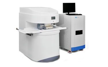
MRI: shake it and see
The MRI machine uses a stronger magnetic field to make the direction of the lines of magnetic force of all water molecules in the body consistent. At this time, the magnetic field of the MRI machine suddenly disappeared, and the magnetic field lines of the water molecules in the human body suddenly returned to the original randomly arranged state. In short, this is equivalent to shaking the water molecules by hand to make them vibrate, then calm down and feel the vibration inside. Therefore, MRI is also called a kind of shock examination.
Which kind of examination is suitable for the four imaging methods?
When seeing a doctor, the doctor usually performs various imaging tests: B-ultrasound, CT, MRI, etc. Many patients will question whether the doctor intends to carry out expensive examinations. In fact, doctors choose different imaging examinations according to different situations.
1. Traumatic bones-take a rough look at the x-rays and carefully scan the CT
For all types of injuries, if it is suspected that the bone has been injured, x-rays are preferred, and the test results are quick and easy to obtain. For further observation, you can choose CT. B-ultrasound and MRI cannot clearly see the bone cortex and medulla, so they are generally not selected.
2. Cervical and lumbar vertebrae-the best option is MRI, the second option is CT
Cervical spondylosis, lumbar disc herniation and other disc diseases require observation of the disc and corresponding nerve roots. In order to better observe these soft tissues, the best choice is MRI. Similarly, MRI is also the first choice for joint, muscle and fat tissue examination.
3. Chest-take a rough look at the x-rays and carefully scan the CT.
X-ray chest radiograph can roughly check the heart, aorta, lungs, pleura, ribs and so on. , And can check whether there is increased lung texture, pulmonary calcification, aortic calcification, etc.
Chest CT examination shows a clearer structure, and its sensitivity and accuracy for chest lesions are better than conventional chest X-rays, especially for the early diagnosis of lung cancer. However, the radiation dose of CT examination is higher than that of X-ray. MRI is very limited in the diagnosis of lung diseases.
4. Abdominal cavity and pelvic cavity-In addition to the intestine, conventional ultrasound examination can be performed
Abdominal organs are greatly affected by breathing, which further affects CT and MRI, while ultrasound is not affected. At the same time, ultrasound has a high diagnostic accuracy for liver, spleen, pancreas, kidney, pelvic organs, etc.
However, the ultrasonic waves are greatly disturbed by the gas. For organs with more gas, such as the intestine, the accuracy of ultrasound diagnosis will be reduced.
5. Cardiac CT is used to exclude coronary heart disease, and ultrasound is used to observe heart function
In routine cardiac structure and function tests, the information provided by cardiac color Doppler ultrasound has been relatively sufficient and simple.
Coronary arteries can be examined with CT, but coronary arterial CT examination has a lot of radiation, which is not suitable for routine physical examination. Although MRI has no electromagnetic radiation, the observation of coronary arteries is not as good as CT. Cardiac MRI is the “gold standard” for evaluating the structure and function of the heart.