 NIUMAG
Small Animal MRI System in Evaluation of Brain Injury in Rats
Small Animal MRI is a powerful non-invasive tool that can be used to detect a wide variety of lesions.
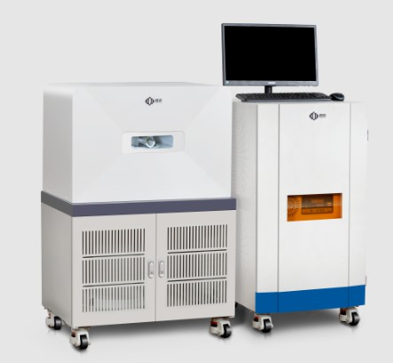
Niumag has developed and produced a new compact, high-performance small animal MRI system that utilizes a low-field NMR design. This small animal MRI system is an application-based approach, to reduce the cost and complexity of traditional systems. The small animal MRI system is mobile and self-shielded and can be placed in most research facilities. No refrigerant or utility supply is required of this small animal MRI. The advantage of this small animal MRI system is that it can easily provide clear 3D digital morphological images of the entire target organ.
In small animal MRI analysis, spin-lattice relaxation time (T1) and spin-spin relaxation time (T2) values are frequently encountered, the normal patterns of which vary by organ, tissue, and fluid . When brain injury was induced in rats, T1 and T2 signals were correspondingly altered due to tissue changes; these T1 and T2-weighted image changes allowed us to detect induced brain injury.
Pilocarpine, a muscarinic cholinergic agonist, is a widely accepted drug for inducing epilepsy and morphological neuronal damage in the rat brain. In this neuronal injury model, clear histological brain injury can be observed in multiple parts of the brain, such as the piriform cortex, lateral dorsal nucleus of the thalamus, hippocampus, and substantia nigra.
Small Animal MRI Analysis
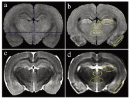
Figure 1 shows T1 and T2 weighted MRI images. Compared with controls, pilocarpine-treated animals showed high T1 signal in the piriform cortex, lateral thalamic nucleus, retroperiventricular nucleus of the thalamus, and posterior hypothalamic nucleus of the brain (Figure 1a and b). In T2-weighted images of pilocarpine-treated animals, compared with controls, low T2 signal was observed in the piriform cortex, corresponding to areas of high T1 signal (Figure 1c and d). The T2 signal intensities of the other 3 areas of high T1 signal were comparable to those in the control group (moderate intensity) (Figures 1c and d).
Applicability of an easy-to-use compact small animal MRI system in the clinical toxicology-pathological examination of pilocarpine-induced rat brain injury. High T1 and low T2 signals showed marked histopathological neuronal damage, although histopathological examination was more sensitive.
The use of small animal MRI systems produced by Niumag is of high value for the study of brain injury.