 NIUMAG
Small Animal Imaging MRI System for Preclinical Research
Small Animal Imaging MRI System
Magnetic Resonance Imaging (MRI) is widely used in preclinical research and drug development. It is a powerful non-invasive method that can be used to evaluate the phenotypes and treatment effects of mouse disease models. In vivo small animals imaging enables longitudinal assessment of tissue changes and phenotype expression in experimental animal models. The NIUMAG small animal imaging MRI System can be used in most conventional laboratories, eliminating the high cost and complexity associated with traditional MRI systems, providing a high-performance experimental platform for in vivo small animal imaging. This article mainly introduces the research applications of the NIUMAG compact, high-performance small animal imaging platform in preclinical toxicology and pathology.
Advantages of Small Animal Imaging MRI
Small Animal Magnetic Imaging (MRI) is a powerful non-invasive method for in vivo evaluation in preclinical research, drug development, and small animal disease models. In preclinical research, in vivo MRI can be used for longitudinal studies to non-invasively monitor the progression, regression, and treatment processes of diseases. Animals do not need to be sacrificed at different time points, allowing imaging of the same animal at different stages of disease development and eliminating individual variations.
So far, the widespread application of MRI technology has been limited by the high procurement costs of traditional superconducting MRI systems and the costs associated with site selection, installation, operation, and routine maintenance of these MRI systems. Additionally, operating superconducting MRI systems poses significant safety concerns and complexity, requiring specialized technical personnel with specific knowledge of MRI physics to operate the instrument and its complex sequences and imaging protocols.
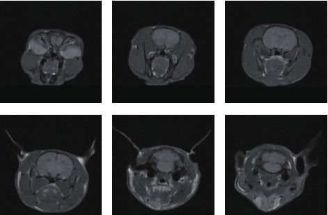
MRI of Mouse Brain
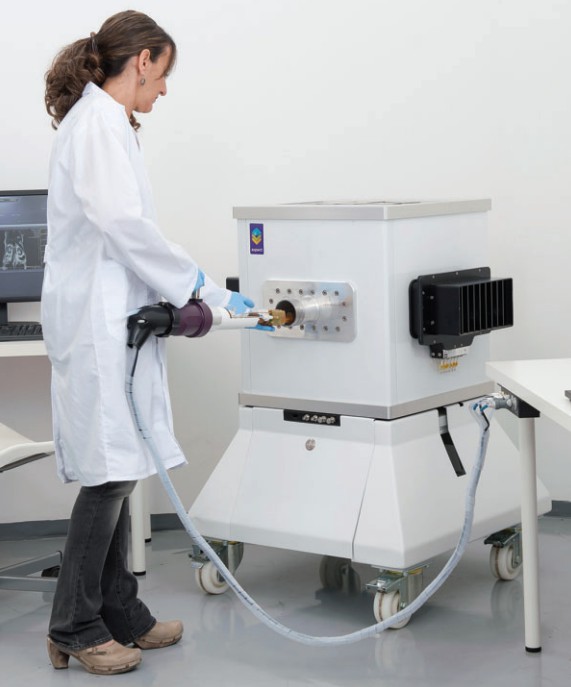
NIUMAG Small Animal Imaging
The NIUMAG compact and high-performance small animal MRI platform is developed based on new magnet design and application software, focusing on in vivo applications for small animals. This system addresses the high cost and complexity of traditional MRI systems, offering user-friendly instrumentation and simplified operation. It is custom-designed for pathologists without a background in MRI, providing high-quality in vivo MRI images of experimental animals and significantly enhancing routine histopathological research in preclinical toxicology and the development of rodent models for human diseases.
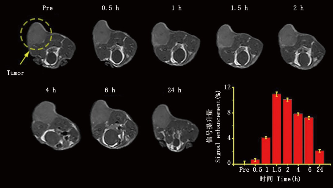
MRI of Subcutaneous Tumor
Unlike superconducting MRI systems, the NIUMAG small animal imaging system is portable and self-shielded, allowing it to be placed in most laboratories or research facilities without the need for special shielded rooms, cryogens, coolants, dedicated electrical supplies, or pipelines.
Furthermore, the system features dedicated software and hardware, as well as pre-programmed protocols and sample handling systems, to facilitate high-throughput imaging of live animals by pathologists. The advantages of this new system include the ability to longitudinally monitor diseases (in vivo MRI) and rapidly acquire magnetic resonance images.
In the past, the use of MRI for small animal imaging was limited to large imaging and research centers that could afford, maintain, and equip expensive superconducting MRI systems, along with supporting complex infrastructures and operating technically challenging software and instruments. These centers have successfully utilized nuclear magnetic resonance imaging (MRI) to produce detailed high-resolution in vivo and ex vivo images while balancing long acquisition times and low sample throughput.
The introduction of the NIUMAG compact small animal imaging system allows researchers and pathologists without prior MRI background and experience to access and effectively utilize the powerful imaging capabilities of MRI without the associated costs, complexity, and safety concerns of superconducting MRI systems.
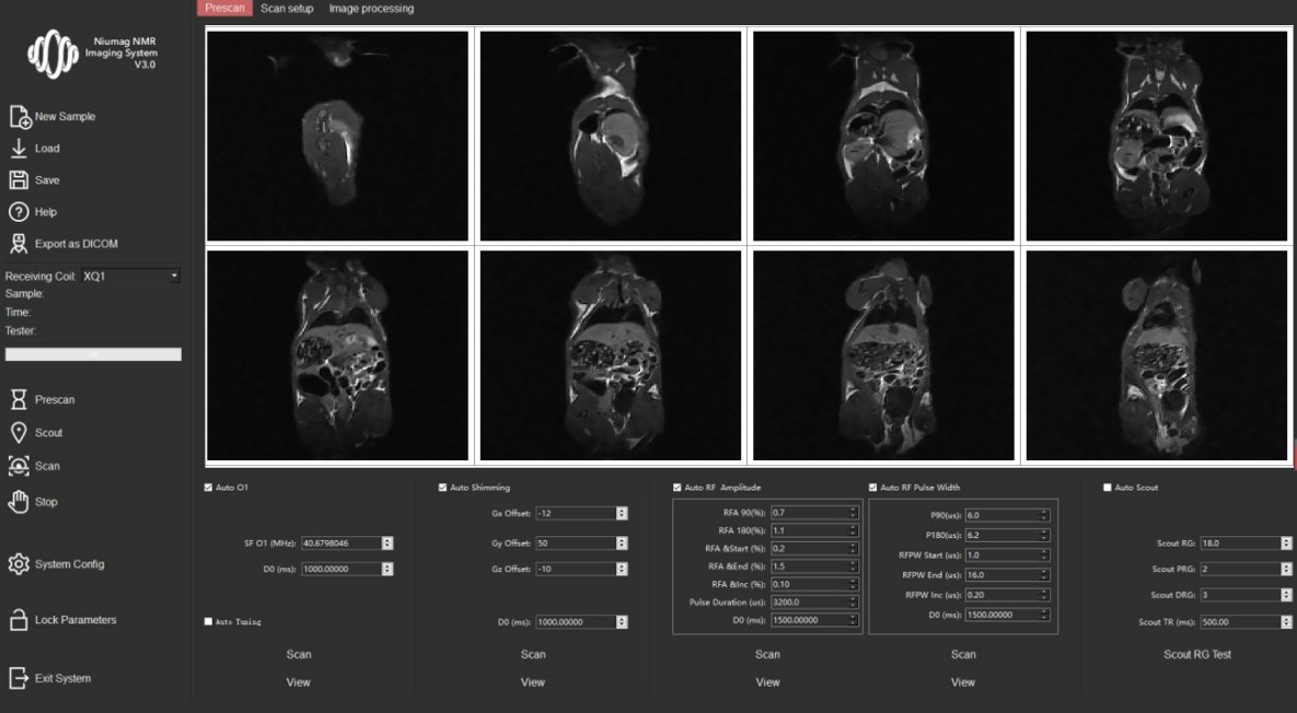
NIUMAG MRI Software Operation
Main Advantages of the NIUMAG Compact Small Animal Imaging (MRI) System:
- High-intensity permanent magnet with a magnetic field strength of up to 1.0T, with no maintenance costs: Reduces procurement and maintenance expenses.
- Compact and small design, easy installation, space-saving, no need for shielded rooms.
- Powerful and well-equipped, reducing the entry barrier for usage.
- Simple operation, fully functional software, optional features such as electrocardiogram monitoring, respiratory gating, and gas anesthesia.
- Exclusive service team from NIUMAG providing professional, high-quality, and prompt technical support services.
- Over 30 sets sold and installed in China.
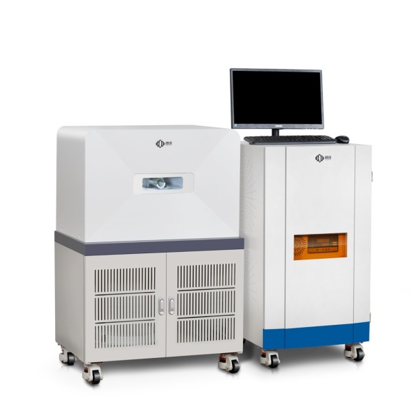
NIUMAG Small Animal Imaging MRI System NM21-060H-I
The NIUMAG compact Small Animal Imaging system provides highly valuable data for researchers by performing multiple imaging sessions on the same animal. It also enables fast imaging, significantly improving experimental efficiency and output. Therefore, the NIUMAG compact small animal imaging system has become an essential tool in the field of life sciences. With continuous technological advancements, this system will continue to gain favor among researchers, bringing us more discoveries and achievements in the future.