NIUMAG
Small Animal imaging Methods
Small Animal Imaging Methods
Small animal imaging refers to the use of various imaging techniques to study and visualize the anatomy, physiology, and pathology of small laboratory animals, typically rodents such as mice and rats. These imaging techniques enable researchers to non-invasively examine and monitor disease progression, evaluate treatment efficacy, and study biological processes in live animals.
Several imaging modalities are commonly employed in small animal imaging:
- Magnetic Resonance Imaging (MRI): MRI uses strong magnetic fields and radio waves to generate detailed images of the internal structures of animals. It provides excellent soft tissue contrast and enables researchers to visualize anatomical features, detect tumors, and study organ function.
- Computed Tomography (CT): CT scanning involves taking multiple X-ray images from different angles and combining them to create detailed cross-sectional images of the animal’s body. CT provides high-resolution images of bones, blood vessels, and organs, and is particularly useful for studying skeletal abnormalities and vascular diseases.
- Positron Emission Tomography (PET): PET imaging utilizes the injection of a radioactive tracer that emits positrons. The emitted positrons collide with electrons, producing gamma rays that are detected by the PET scanner. This technique allows researchers to track and quantify metabolic processes, study specific molecular targets, and assess the distribution of radiolabeled drugs in the animal’s body.
- Single-Photon Emission Computed Tomography (SPECT): SPECT imaging is similar to PET, but it uses different radioactive tracers that emit single photons. SPECT provides 3D images of radiotracer distribution in the body, enabling the assessment of physiological processes and the detection of specific molecular targets.
- Optical Imaging: Optical imaging techniques, such as bioluminescence imaging and fluorescence imaging, utilize light to visualize specific molecules or cells within the animal. Bioluminescence imaging involves the detection of light emitted by bioluminescent reporters, while fluorescence imaging uses fluorescent probes that emit light when excited by a specific wavelength. These techniques are widely used for tracking gene expression, studying cellular processes, and monitoring tumor growth in small animals.
- Ultrasound Imaging: Ultrasound uses high-frequency sound waves to create images of internal structures in real-time. It is particularly useful for imaging the cardiovascular system, visualizing tumors, and guiding interventions in small animals.
Small animal imaging plays a crucial role in preclinical research, allowing researchers to gain insights into disease mechanisms, evaluate novel therapeutics, and develop new diagnostic tools. It enables the study of disease progression in real-time, reduces the number of animals required for experiments, and provides a non-invasive alternative to traditional invasive methods.
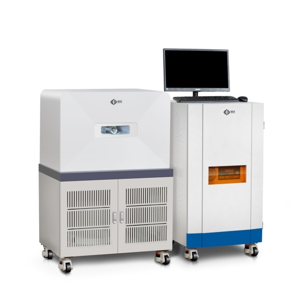
Small Animal Imaging by Low Field NMR
Low-field NMR (Nuclear Magnetic Resonance) refers to the use of NMR technology at lower magnetic field strengths compared to conventional clinical MRI systems. While high-field MRI systems, typically operating at 1.5 Tesla (T) or higher, are commonly used for human imaging, low-field NMR systems operate at field strengths ranging from a few milliTesla (mT) to several tens of milliTesla (mT).
In the context of small animal imaging, low-field NMR can be utilized to obtain anatomical and functional information from small laboratory animals. Here are a few points to consider regarding the application of low-field NMR in small animal imaging:
Resolution and Sensitivity: Low-field NMR systems generally provide lower resolution compared to high-field MRI. The spatial resolution of the images may be limited due to the lower magnetic field strength. However, for certain applications in small animal research, such as monitoring disease progression or assessing treatment response, the achievable resolution may still be sufficient.
Cost and Accessibility: Low-field NMR systems are typically less expensive and easier to maintain compared to high-field MRI systems. This can make them more accessible to researchers with limited resources or those working in small laboratory settings.
Safety Considerations: Low-field NMR systems generally have lower magnetic field strengths, which may result in reduced safety concerns compared to high-field MRI systems. However, it is still important to follow safety guidelines and ensure the proper handling of the animals during imaging.
Applications: Low-field NMR can be employed in various small animal imaging applications. It can provide anatomical information, such as organ morphology, and can be used to monitor disease progression in longitudinal studies. Additionally, low-field NMR can be combined with contrast agents or specific pulse sequences to extract functional information, such as blood flow or tissue perfusion, in small animals.
Advantages and Limitations: The use of low-field NMR in small animal imaging offers some advantages, such as cost-effectiveness, ease of use, and reduced safety concerns. However, the lower signal-to-noise ratio (SNR) compared to high-field MRI can limit the detection sensitivity and overall image quality. Additionally, the availability of specialized imaging sequences and hardware configurations may be more limited at lower magnetic field strengths.
In summary, low-field NMR can be a valuable tool for small animal imaging, offering cost-effective and accessible imaging capabilities. While it may have limitations in terms of resolution and sensitivity compared to high-field MRI, it can still provide valuable anatomical and functional information for preclinical research applications.
NIUMAG NM21-060H-I is designed for in-vivo MRI observation of animals. It is a permanent magnet system that provides high contrast images and has an intuitive user interface. As a powerful and non-destructive MRI instrument, this system is widely used in life sciences for in-vivo study of tissue structures and contrast agent dispersion. The Niumag small animal MRI system is robust with low operating and maintenance costs.