 NIUMAG
Obese Mice NMR Imaging
Obese Mice
Obese mice are a commonly used animal model in scientific research to study the biology and underlying mechanisms of obesity. These mice are genetically or diet-induced to develop obesity, which is characterized by excessive accumulation of body fat and weight gain.
Obese mice are used to study various aspects of obesity, including its causes, effects on metabolism and physiology, and potential treatments. For example, scientists use obese mice to study the role of genes, hormones, and environmental factors in the development of obesity. They also use these mice to investigate the metabolic changes associated with obesity, such as insulin resistance and altered lipid metabolism.
In addition, obese mice are used to test potential treatments for obesity, including drugs, dietary interventions, and exercise. These interventions can help identify potential targets for therapy and provide insight into the mechanisms of action of these treatments.
Obese Mice NMR
In scientific research, NMR (Nuclear Magnetic Resonance) spectroscopy is a commonly used technique for studying the composition and structure of molecules, including those found in biological samples.
Regarding obese mice, NMR can be used to study changes in the metabolic profile of these animals compared to lean mice. For example, NMR can be used to analyze the concentrations of various metabolites in biological samples such as blood, urine, or tissue extracts, which can provide insights into the underlying metabolic changes associated with obesity.
NMR can also be used to study the effects of interventions such as drugs or dietary changes on the metabolic profile of obese mice, which can help identify potential therapeutic targets or interventions for obesity.
Overall, NMR is a useful tool for studying the metabolic changes associated with obesity in mice and other animal models, and can provide important insights into the underlying biology of obesity and potential therapeutic targets for this condition.
Obese Mice Imaging
Observation and comparison of 25g normal mouse/lung tumor mouse tumor
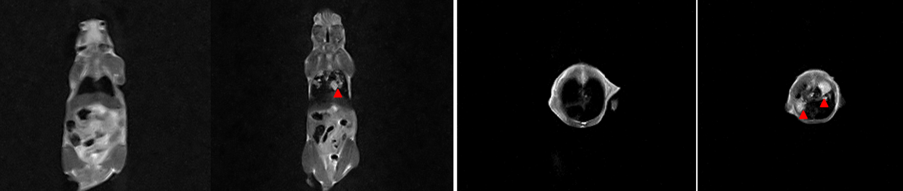
The main body of the lung is a cavity, and the grayscale image in the magnetic resonance imaging generally shows a black background with no signal. If there is a signal, it can be speculated to be a suspected lesion tissue.
Orthotopic Tumor Imaging in Mouse Liver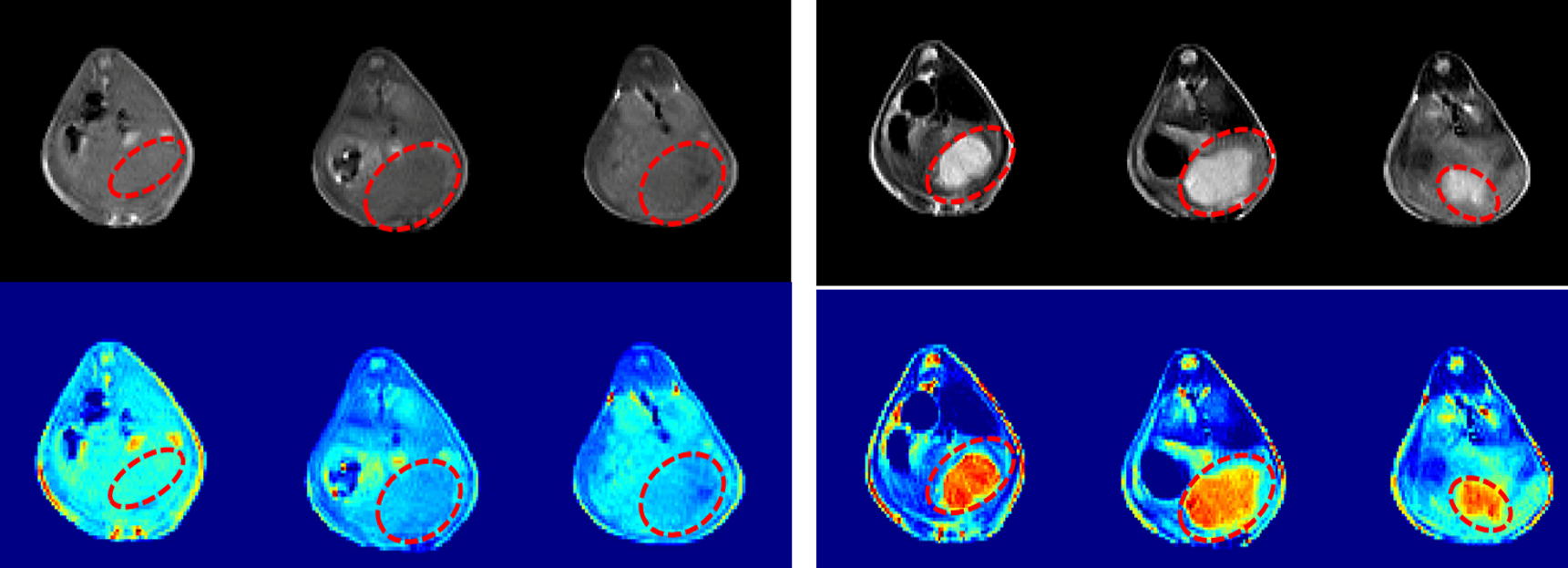
Compared with normal tissue, the components of the tumor have longer relaxation, so the signal is prominent in the T2-weighted image.
Imaging comparison before and after contrast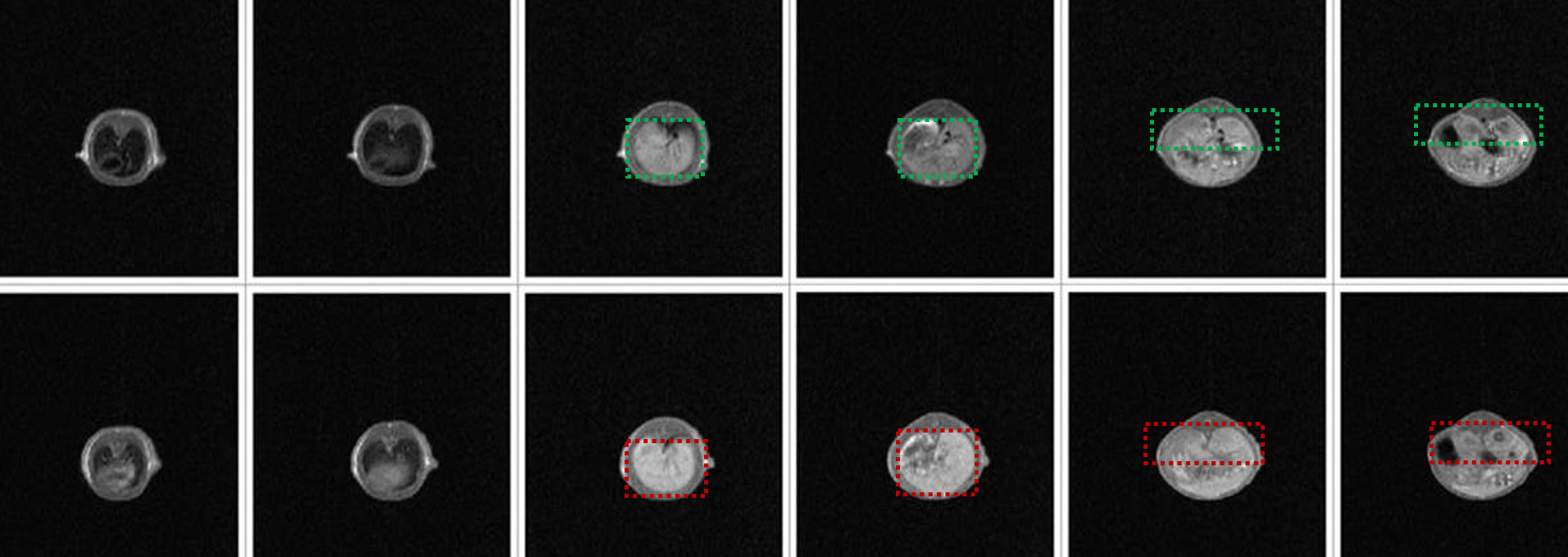
Comparison of T1-weighted imaging before and after contrast agent injection. The gray value of the liver (Fig. 3/4) changes significantly before and after contrast, and the kidney (Fig. 5/6) changes slightly, indicating that the contrast material is better enriched in the liver.
Imaging of the metabolic process of contrast agents in vivo
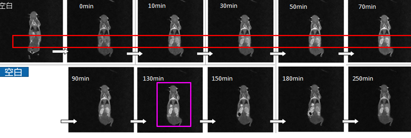
Imaging changes in mice at 0-250min after contrast agent injection
It can be seen from the imaging image that the metabolism time of this contrast agent in the kidney exceeds 250 minutes, and reaches the peak value at 130 minutes.
Imaging of Metabolic Processes in Subcutaneous Tumors after Contrast Imaging
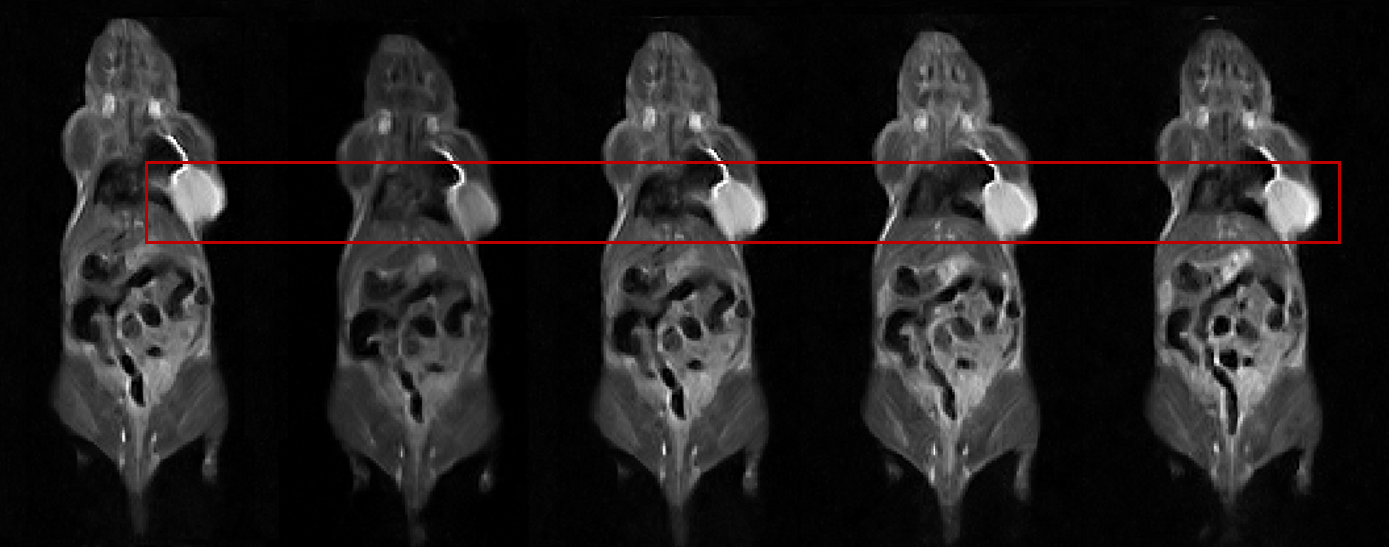
MRI T2-weighted imaging evaluates the metabolic process of the tumor after injection of contrast medium in mice. Imaging is performed at different time periods after injection of contrast medium. MRI corresponds to before injection, 0.5h after injection, 1h after injection, 1.5h after injection, and 2h after injection.
Overall, obese mice are a valuable tool in scientific research to study the biology of obesity and develop new treatments for this condition. However, it is important to note that animal models may not perfectly replicate the human condition, and results from animal studies should be interpreted with caution before translating to clinical practice.