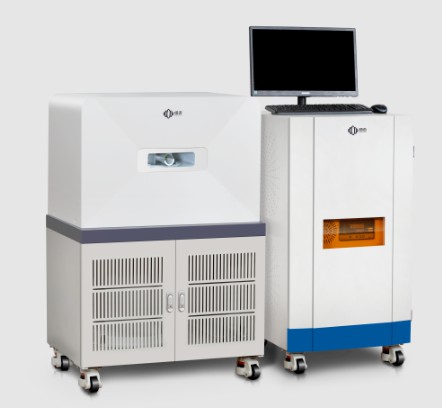
NIUMAG
Mouse MRI
Mouse MRI Background
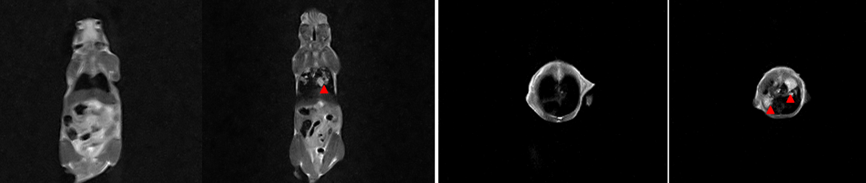
Mouse MRI (Magnetic Resonance Imaging) is a medical imaging technique that uses a powerful magnetic field, radio waves, and a computer to produce detailed images of the internal structures of a mouse’s body. Mouse MRI is a non-invasive method of imaging that can help researchers to understand the structure and function of organs, tissues, and other structures in living mice.
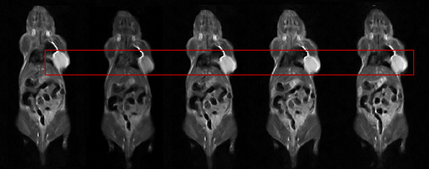
Mouse MRI can be used to image a wide range of structures in mice, including the brain, heart, lungs, liver, kidneys, and other organs. It can also be used to study disease processes, monitor the effects of treatments, and assess the effectiveness of drugs.
Mouse MRI can be performed using a variety of techniques, including T1-weighted imaging, T2-weighted imaging, diffusion-weighted imaging, and functional MRI. The choice of technique depends on the specific research question being investigated.
Mouse MRI is an important tool for researchers studying diseases and conditions that affect mice, such as cancer, Alzheimer’s disease, Parkinson’s disease, and cardiovascular disease. It can also be used to study the effects of environmental toxins, drugs, and other treatments on the mouse body.
Mouse Image Methods
There are several methods that can be used to obtain images of mice for research purposes, including:
- Micro-Computed Tomography (micro-CT): This imaging method uses X-rays to create high-resolution 3D images of the mouse body. It is particularly useful for studying bone structure, but can also be used to visualize other tissues and organs.
- Magnetic Resonance Imaging (MRI): As mentioned earlier, MRI uses a strong magnetic field and radio waves to create detailed images of the internal structures of the mouse body. It is non-invasive and can be used to study a wide range of tissues and organs.
- Optical Imaging: This method uses light to create images of tissues and organs, and can be used to study a range of biological processes, such as gene expression and blood flow. Different types of optical imaging include bioluminescence imaging (BLI) and fluorescence imaging (FLI).
- Ultrasound: This method uses sound waves to create images of internal organs and tissues, and is particularly useful for studying cardiovascular function and blood flow.
- Positron Emission Tomography (PET): This method involves the injection of a radioactive tracer into the mouse body, which emits positrons that are detected by a PET scanner. It can be used to study a range of biological processes, such as metabolism and receptor binding.
- Single Photon Emission Computed Tomography (SPECT): This method is similar to PET, but uses a different type of radioactive tracer. It can also be used to study a range of biological processes.
These methods can be used alone or in combination with each other to obtain a comprehensive understanding of the mouse anatomy and biology, and to study disease processes and treatment efficacy.
Mouse MRI by Low Field NMR
Low field NMR (Nuclear Magnetic Resonance) is a type of MRI that operates at a lower magnetic field strength than traditional clinical MRI machines. It is a relatively new technology that is being developed for preclinical research, including studies involving mice.
Mouse MRI by Low Field NMR
Low field NMR is an attractive technology for mouse MRI because it is relatively low cost and does not require a specialized facility to operate. It can also be used to study a wide range of mouse models and tissues, including the brain, liver, kidneys, and other organs.
One of the main advantages of low field NMR is that it can be used to study samples in their natural state, without the need for invasive procedures. This means that researchers can study the natural structure and function of mouse tissues and organs, which is important for understanding disease processes and developing new treatments.
However, low field NMR has some limitations compared to high field MRI machines. For example, the resolution of the images produced by low field NMR is generally lower, which can make it more difficult to study fine details in the mouse anatomy. Additionally, low field NMR may not be suitable for studying certain types of tissues or disease processes.
Low field NMR for Mouse MRI Advantages:
- Lower cost: Low field NMR machines are generally less expensive to purchase and maintain than high field MRI machines, which makes them more accessible to researchers with limited budgets.
- Lower magnetic field strength: Low field NMR machines operate at lower magnetic field strengths than high field MRI machines. This means that they are less affected by artifacts caused by movement or other sources of noise, which can improve image quality.
- Reduced sensitivity to metal artifacts: High field MRI machines are sensitive to metal artifacts, which can interfere with image quality. Low field NMR machines are less sensitive to these artifacts, which makes them useful for studying tissues and organs that contain metal implants, such as orthopedic implants.
- Reduced motion artifacts: The lower magnetic field strength of low field NMR machines reduces the occurrence of motion artifacts in images, which can be particularly important for studying the mouse heart and other organs that are prone to motion.
- Increased tissue contrast: Low field NMR machines can provide increased tissue contrast in some cases, which can be useful for studying certain tissues and organs that are difficult to visualize with high field MRI machines.
Overall, low field NMR is a promising technology for mouse MRI that offers several advantages over traditional high field MRI machines. It may be particularly useful for studying mouse models of disease and for preclinical research applications where cost and accessibility are important factors.