NIUMAG
Low Field NMR Technique in Contrast Agent and Animal Imaging
MRI is widely used clinically because of its non-invasive, rapid, high-resolution, and high-contrast features. Especially in the diagnosis of tumors, the technology has become an indispensable and important basis for the early diagnosis of primary tumors and tumor metastasis by using the structural and functional images obtained from the physical properties of the diseased tissue and normal tissue. The formation of tumor is a complex process of long-term, multi-factor control, multi-step, multi-gene mutation. Most malignancies are of monoclonal origin and exhibit uncontrolled growth. Clinically, when a considerable number of patients seek medical treatment, the disease has entered the middle or late stage, and the optimal treatment time has been lost, which is one of the reasons for the high tumor mortality rate. Although MRI has the above advantages, it cannot meet the requirements of early diagnosis of tumors due to its low sensitivity. This is because early-stage tumors and normal tissues have small differences in physical properties (eg, T1 and T2), and this small difference in physical properties is not sufficient to generate image contrast between tumor and normal tissues. In order to solve this problem, people use MRI contrast agents to enhance the contrast of tumor and normal tissue images to facilitate the early diagnosis of tumors.
Principles of MRI Contrast Agents
The MRI signal of hydrogen nuclei is the MRI signal source of various tissues. MRI contrast agents do not produce signals, but their role is to change the relaxation time of the hydrogen nuclei system inside the tissue. Contrast with surrounding tissue. MRI signal intensity is related to physical and chemical parameters, such as proton density spin-lattice relaxation time T1, spin-spin relaxation time T2. The T1 and T2 parameters control the contrast intensity of the imaging. Hydrogen proton density changes very little in soft tissue, so T1-weighted imaging and T2-weighted imaging are used in diagnosis. The function of a contrast agent depends on its concentration in the tissue and on the density and movement of protons in the tissue.
MRI contrast agents must be magnetic substances that can interact magnetically with hydrogen nuclei. The contrast agent mainly changes the signal intensity by affecting T1 and T2. According to the principle, contrast agents can be divided into T1-type preparations and T2-type preparations. T1-like preparations increase the intensity of the signal on T1-weighted imaging. T2-type preparations reduced signal intensity on T2-weighted imaging. Which type of contrast agent is used clinically depends on the characteristics of the tissue. The process of T1 shortening requires the direct interaction of hydrogen protons with the magnetic part of the contrast agent, that is, the hydrogen nucleus of the water molecule should be as close as possible to the magnetic particles to achieve relaxation enhancement. For example, the T1 enhancement effect of liposome-encapsulated Gd-DTPA is weaker than that of Gd-DTPA at the same concentration, because the liposome restricts the access of external water molecules to Gd-DTPA. The T2 shortening process is a long-range effect that interferes with T2 through the inhomogeneity of the local magnetic environment of the T2 preparation. Encapsulation of T2-like formulations into liposomes results in enhanced T2 relaxation because aggregation of the liposomes produces greater changes in the local magnetic environment.
The relationship between T1 and T2 values in tissue and contrast agent concentration is as follows:
![]()
R1 and R2 are the relaxation rates of the contrast agent, which describe the effect of the contrast agent on T1 and T2 in the tissue, and [c] is the concentration of the contrast agent evenly distributed in the tissue. Formulas (1) and (2) show a simple linear relationship between the reciprocals of T1 and T2 and the concentration of the contrast agent. R1 and R2 depend on the structure of the contrast agent. For T1 type preparations, it mainly depends on the proximity of the contrast agent to water molecules, and for T2 type preparations, it depends on the concentration of magnetic substances.
MRI tumor diagnosis
In 1971, Damadian found that the T1 and T2 values of the relaxation time of water molecule protons in tumor tissue are larger than those in normal tissue. , chest and abdomen images. Since then, nuclear magnetic resonance imaging technology has been highly valued by the scientific community for its outstanding advantages such as no radiation damage, no destructiveness, no reagent invasion, and the ability to systematically study living and dynamic processes from the molecular level to the whole organ. Rapidly, this new medical imaging diagnosis technology has been rapidly popularized in medical centers and large and medium-sized hospitals in various countries and has become an important tool for clinical diagnosis.
The principle of MRI diagnosis technology is mainly to use water protons in different tissues of the organism to generate different resonance signals under the influence of an external magnetic field for imaging. The strength of the signal depends on the water content in the tissue and the relaxation time of protons in water molecules. It can effectively detect tissue necrosis, ischemia and various malignant lesions (such as tumors) for early diagnosis.
Show results
The instrument uses a small animal MRI system, and the images are T1-weighted cross-section and coronal plane. The sampling parameters are as follows: FOV=100mm×100mm, TR=400ms, TE=19ms, slice thickness 3.5mm, slice interval 1mm, accumulation times 16, the size of K space is 192×256. The imaging results showed that after the injection of the contrast agent, the rat heart and liver became brighter, and gradually became darker with the prolongation of metabolic time.
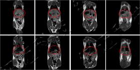
MRI coronal phase before and after angiography in rats
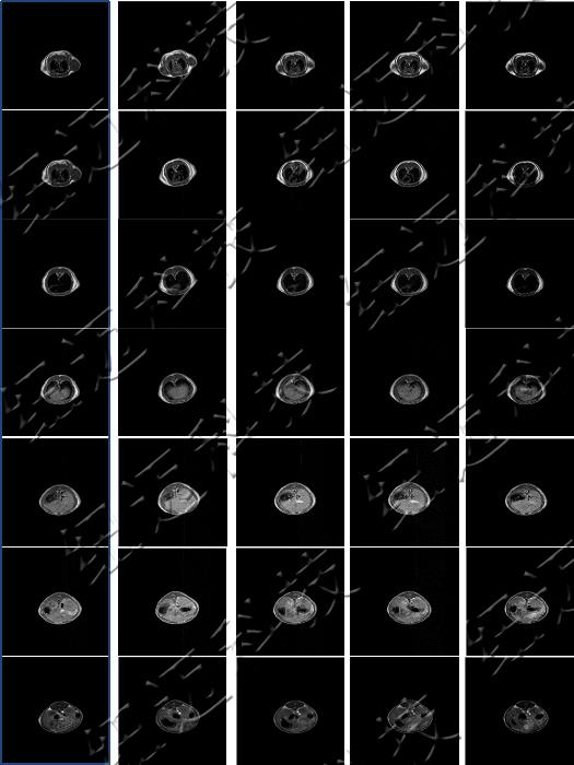
MRI cross-section of contrast agent metabolism in rats